
Selling fast!
Get yours while you can.
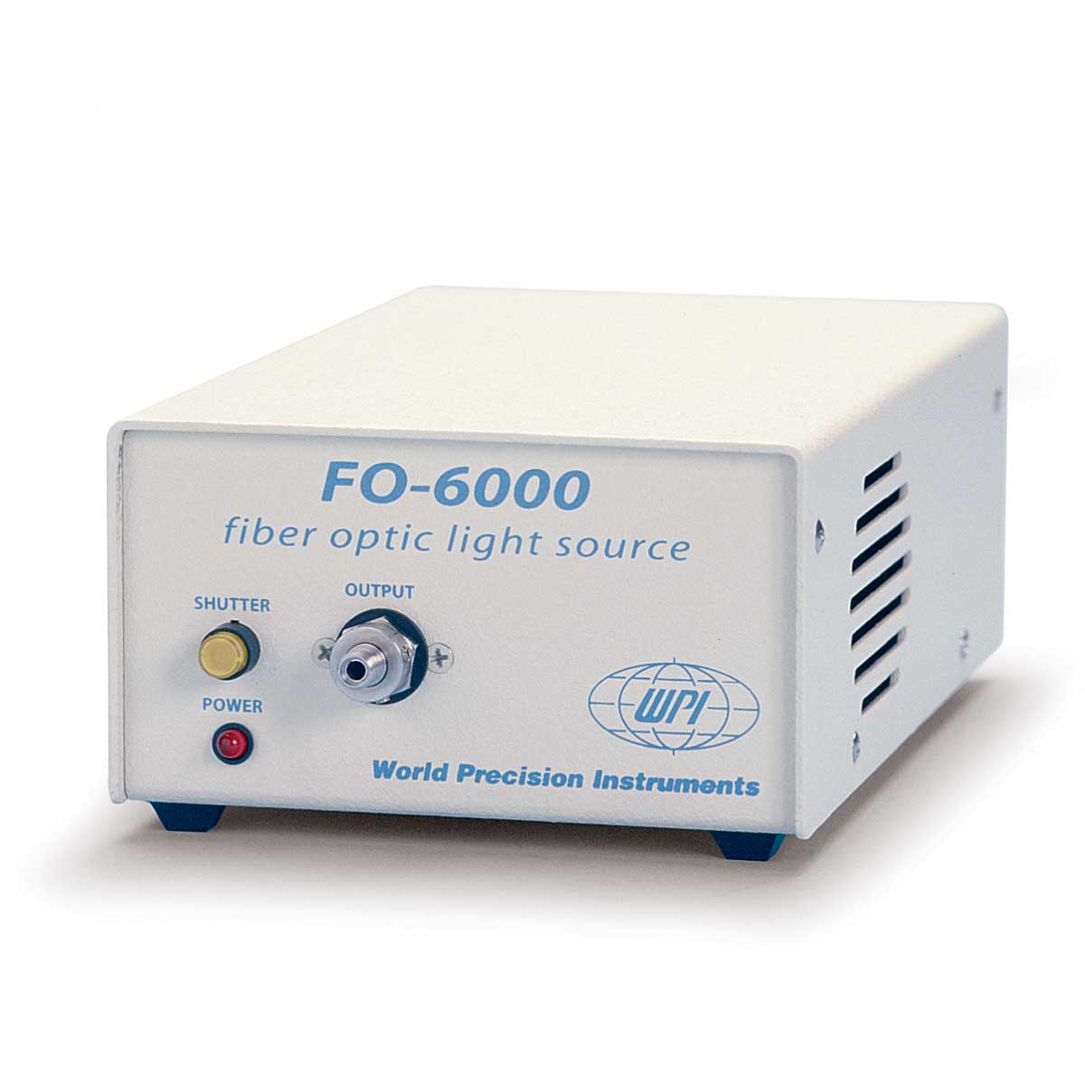
FO-6000
Couldn't load pickup availability
Prices valid in USA, Canada, and PR only.
Click here to view the current Data Sheet.


56200

89575


Multiple SKUs


503847

503848


Multiple SKUs

CUV2061-1

CUV2062-1

CUV2071-1


D4H

Multiple SKUs

TIDAS-D2


Multiple SKUs