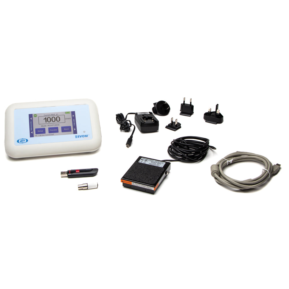
EVOM™ Manual Meter for TEER Measurement

Skip to product information
1 of 6
Regular price $4,500
Sale price $4,500 Regular price

EVM-MT-03-02


Prices valid in USA, Canada, and PR only.

The EVOM™ Manual replaces all previously manufactured WPI manual TEER meters, including the EVOM3, EVOM2, and MilliCell® ERS-2, which have now been discontinued.

This next-generation manual TEER meter features automatic data logging for streamlined data collection. Its low-noise design ensures greater resolution and accuracy, while the automatic 20X sample averaging enhances measurement stability and precision. Select from adjustable fixed measurement currents (2, 4, or 10 μA) to suit your protocol. The auto-ranging resistance measurements span from 1 Ω to 100,000 Ω, with three fixed current range options for added flexibility. 

Engineered for reliability, the low-current, low-voltage design prevents metal ion transport and delivers fast resistance stabilization for low-level readings under 200 Ω, with a resolution as fine as 0.1 Ω. The ergonomic tilt stand minimizes glare for comfortable use, while automatic plate indexing supports operation with or without control well subtraction for resistance and potential difference (PD) measurements. Enjoy continuous data logging via USB flash drive, which is compatible with PC, Mac, and Linux, ensuring seamless integration into your workflow.

NEW FEATURE:  In addition to the existing capability of data storage on a USB flash drive, the new version of the EVOM™ Manual now provides an option for a more secured mode of data transfer using a Windows® companion application.

ACCESSORIES


Details

TEER measurement device with automatic data logging

evom 360 view

Features

  • Low noise design offers greater resolution and accuracy in your TEER measurement protocol
  • Automatic 20X sample averaging improves accuracy and stability
  • Adjustable fixed measurement currents (2, 4 or 10 μA)
  • Resistance auto ranging from 1 Ω to 100,000 Ω or with three fixed current ranges
  • Reliable low current, low voltage design prevents metal ion transport
  • Fast resistance stabilization on low levels under 200 Ω with resolution to 0.1 Ω
  • Ergonomic tilt stand for low glare operation
  • Graphical display of popular plates (6, 12, 24, 96) for trend analysis
  • Display shows the most recent set of parameters
  • Automatic plate indexing operation with or without control well subtraction for resistance and potential difference (PD) measurements
  • Continuous data logging via USB (PC, Mac, Linux)
  • Saves date stamped data to a spreadsheet readable file on a USB drive
  • Upgradable firmware
  • Upgrade of the EVOM3, in the trusted family of the EVOM™ brand of TEER measurement products

Benefits of the EVOM™ Manual TEER Measurement Device

  • Eliminates errors and reduces experimental processing time
  • Auto data logging eliminates the need to track data by hand
  • The small footprint allows more bench space
  • Easy calibration and verification
  • Footswitch for hands-free recording
  • Prevent data loss with auto save and data recovery when battery is low
  • Easy TEER value calculation by applying a unit area formula to the resistance
  • Premium Warranty Available

Applications for TEER Assays

  • Measure epithelial or endothelial tissues for confluence, TEER and potential difference
  • Permeability, conductance and drug studies
  • Continuous digital monitoring of a target membrane
  • Common studies
  • Blood-brain barrier transport
  • Lung epithelial tissue studies
  • Intestinal tissue studies
  • Skin studies

EVOM™ Manual For TEER Measurement

The EVOM™ Manual delivers improved workflow efficiency, more stable and repeatable measurements versus traditional Trans Epithelial Electrical Resistance (TEER) meters. Providing you with vital feedback during experiment measurements, the EVOM™ Manual’s large screen offers a range of informational views. The new graphical displays for trend analysis and measurement values helps scientists deliver simple, stepwise methodology during experimental measurements. The touch screen interface provides you with an intuitive, easy-to-use menu for configuration.

Eliminating the need to log data by hand, the EVOM™ Manual writes the resistance or voltage information to a USB drive in CSV format for easy transfer to spreadsheets and data analysis programs. When used with the footswitch it enables hands-free recording of measurements.

At the heart of the EVOM™ Manual is our latest processor and circuitry, providing users with quick, easy and reliable readings due to its fast stabilization, automatic twenty times sampling average and low noise design. The auto ranging resistance feature allows for fast resistance measurements, and an over-range display feature eliminates false readings. The EVOM™ Manual has adjustable current levels in three fixed ranges with two lower ranges for sensitive membranes and high resistance ranges up to 100 KΩ.

How Does the EVOM™ Manual Work?

Confluence of a cellular monolayer is determined by an increase or a plateau in tissue resistance detected using the unique electronic circuit of the EVOM™ Manual and the new STX4 electrode. The EVOM™ Manual qualitatively measures cell monolayer health and quantitatively measures cell confluence. The EVOM™ Manual produces a low AC current that avoids electrode metal deposits and adverse effects on tissues which can otherwise be caused by higher DC currents. The EVOM™ Manual uses low current and voltages and is designed for non-destructive testing for epithelial monolayer confluence in cell cultures. In addition, resistance readings are unaffected by membrane capacitance or membrane voltage. The accuracy and repeatability of the EVOM™ Manual-STX4 system makes this instrument ideal for permeability, PD and other detailed membrane studies.

Electrodes For TEER (Epithelial) Measurement

 Part # Descriptions
EVM-EL-03-03-01 STX4 EVOM™ Electrode with Removable Blades for TEER in 6 mm Inserts
STX2* Replacement Electrode Set (Requires 99672 for use with the EVOM3 or EVOM™ Manual)
STX3* Adjustable electrode set for shallow wells, 5-9 mm depth
3993* 2 mm Adapter for EVOM2

*(Requires 99672 for use with the EVOM3 or EVOM™ Manual)

ENDOHM Chambers For Endothelial/Epithelial Measurement

NEW EndOhm chambers include the EVOM™ Manual cable 99916.

 Part # Descriptions
EVM-EL-03-01-01 ENDOHM-6 EVOM™ Electrode for TEER in 6 mm Insert
EVM-EL-03-01-02 ENDOHM-12 EVOM™ Electrode for TEER in 12 mm Insert
EVM-EL-03-01-03 ENDOHM-24 EVOM™ Electrode for TEER in 24 mm Insert  

TEER measurement techniques for in vitro barrier model systems

EVOM™ TEER Measurement Basic Working Principle

What is TEER?

TEER, Trans Epithelial Electrical Resistance, monitures the electical resistance of a cellular layer is the inverse presentation of the electrical conductance through the cellular layer. A high TEER value of the cellular layer is indicative of an intact cellular monolayer and suggests low or restricted permeability of ions and molecules (i.e., low conductance). Similarly, a decrease in the TEER value suggests a compromised barrier function and indicates increased permeability. Tissue permeability studies require a confluent cellular layer, and TEER measurement is gennerally used to confirm the formation of a confluent monolayer. 

Basic working principle of EVOM to measure TEER

Initially, 24 hours after cell seeding transwell, TEER values are generally low, because the current passes can pass easily between the cells. Over time, the cells multiply and start covering the gaps. Finally, a confluent cellular monolayer is formed. At that point the permeable membrane is fully covered with cells and does not allow easy passage of electrical current. This results in a high TEER value.

TEER Value Calculation

TEER is a normalized value of resistance per 1 square centimeter of unit area. To compute TEER, multiply the measured resistance by the surface area listed below.

For example, a 6.6 mm insert measures 1707 Ω, the TEER is 1707 Ω * 0.331 or 505 Ω.

• 6 well plate (24 mm inserts) 4.53 cm2

• 12 well plate (12 mm inserts) 1.13 cm2

• 24 well plate (6.5 mm inserts) 0.3316 cm2

• 96 well plate (4.3 mm inserts) 0.145 cm2


Resources

Manuals

Documents

Software Downloads

Videos

FAQs


Specifications

This unit conforms to the following specifications:

Type Descriptions
 Tissue Sampling Frequency  12.5 Hz
 Sample Averaging  20 samples per second
 Resistance Ranges
  •  0 to 10,000 Ω
  •  0 to 50,000 Ω
  •  0 to 100,000 Ω +5%
 Auto Mode  1 to 100,000 Ω auto current 2 μA, 4 μA, 10 μA
 Resistance Resolution  0.1 Ω (under 200 Ω); 1 Ω (over 200 Ω)
 Resistance Accuracy
  •  0.1 Ω (under 200 Ω), 1 Ω (over 200 Ω) 0.1%
  •  100,000 Ω ± 2 μA (to 105 KΩ)
 Voltage Resolution  0.001 V, 0.1 mV
 Accuracy Resistance  0.1 Ω (200 Ω); 1 Ω (above 200 Ω)
 Accuracy voltage  ± 0.1 mV
 Current Levels
  • 10,000 Ω ±10 μA
  • 50,000 Ω ± 4 μA
  • 100,000 Ω ± 2 μA
  • Auto mode 1 to 100,000 Ω auto current 2 μA, 4 μA, 10 μA
 Display Update Rate  0.5 seconds
 Battery  3.7V Li-ion 2500 mAh**
 Charging Period  5.5 hours (power off); 6 hours (run time)
 Charge Current  200 mA
 Power Consumption  ~250 mA
 Certifications  CE

** mAH means milliamp hours.

System Components

What is included with the EVOM™ Manual QTY
evm-mt-03-01 : EVOM™ Manual Epithelial Volt Ohm Meter  1
300749 : USB drive 32 GB (Used for storage. Also contains a Python 3.8 program for continuous digital monitoring of a target insert).  1
503535 : USB cable  1
99673 : Calibration kit, 1000Ω Test Resistor  1
803025 : A/C power cord and charger  1
13142 : Foot switch  1

NOTE: A 99672 EVOM2 to EVOM Manual Electrode Adapter is sold separately. The STX2, STX3 and all STX100s require the use of this adapter with the EVOM3 or EVOM Manual.

FAQs

Why would I want to use the blank function?

The blank feature is used when you want to subtract out any measurement that is not from the membrane, such as the electrode and fluid resistances.

Does the EVOM Manual system automatically calculate TEER?

No, TEER measurement requires an area calculation. To compute TEER, multiply the measured resistance by the appropriate surface area (below). For example, a 12 mm insert measures 565 Ω, the TEER is 565 Ω × 1.13 cm2 = 638.5 Ω- cm2. Here are the surface areas generally applicable to different transwell/insert formats: 6 well plate (24 mm inserts) 4.52 cm2, 12 well plate (12 mm inserts) 1.13 cm2, 24 well plate (6.5 mm inserts) 0.33 cm2, 96 well plate (4.3 mm inserts) 0.14 cm2. For Automated TEER Measurements, consider using the WPI REMS Automated TEER Measurement System.

EVOM Manual data is stored automatically when the last well is reached. How do I store the data when I only want to measure 8 of 96 wells?

Clear any data in memory by opening settings, store menu then press new plate, that will clear any prior readings. Return to the main screen, open the preview screen, select each well to measure (the selection turns green), place the electrode, then measure. When you’re done measuring the selected wells, open the settings, press the store screen menu, then press store new to save the plate data to the USB drive.

How should you store the EVOM Manual and electrodes if they will be exposed to UV light in a laminar hood for extended periods of time?

Take the EVOM™ Manual out of the laminar hood after use. Next time, turn on the UV inside the hood. Once the hood is disinfected by UV, turn off UV, next spray 70-100% ethanol or isopropanol onto a paper towel and wipe the EVOM™ Manual. Do not spray alcohol directly onto EVOM™ Manual.

Can increasing or changing sample liquid volumes change my resistance values?

You can expect to see a change of raw resistance values. However, you subtract the blank values (blank Transwell with no cells) from the sample values (Transwell with cells). This way, you subtract the blank value with increased volume from samples with increased volume. Thus, any change of resistance contributed by increased volume is omitted. Consistently use the same volumes for all your samples in an experiment.

Why do my readings appear to be drifting?

Drift–Readings that continuously increase or decrease significantly (either voltage or resistance) over time. Example: At 1000 Ω, the reading is increasing 100 Ω/ minute. (A drift of 10 Ω/minute is acceptable.) Excessive drift may be caused by changes in the pH or temperature, or the electrode needs cleaning.

There are a few things that can be a possible cause of this drift.

  • Ensure that the electrodes are fully immersed in culture media solution, and the fluid temperature in the plate is consistent by equilibrating at room temperature or using a plate warmer.
  • Another common cause of drift is the electrode tips may have deposits of cell culture media constituents
  • The electrode (tips) requires enzymatic cleaning (Tergazyme or Enzol) periodically up to 1x per week depending on the use.
  • Handheld electrodes also must be kept as motionless as possible during a measurement.
  • Excessive movement will cause the measurement to fluctuate.
  • Additionally, in a 5% CO2 environment, a loss of CO2 causes the media pH to change, and the resistance reading may change. This is mainly applicable in the context of continuous measurement for an extended period (hours, as compared to a few minutes).

Can you suggest some experimental parameters that can be controlled to obtain consistent TEER results?

Temperature is known to affect TEER values. We recommend that you maintain a consistent temperature to obtain consistent values. Since the readings are obtained in cell culture media/ buffer. We recommend that you use a water bath with a fixed temperature to warm the media/buffer to be used during the experiment. A consistent media/buffer temperature ensures a consistent experimental condition. We recommend taking the well plate, containing cells grown on culture inserts, out of the incubator for at least 20 minutes to stabilize the well plate at room temperature before making measurements. 

If you are using an EndOhm chamber, make sure you maintain the same fixed distance between the top and bottom electrodes to obtain consistent read outs. If you are using a chop-stick electrode (STX2), try to hold it in a vertical position while obtaining results. Consistency in maintaining the same holding position of the chop-stick electrodes while performing an experiment is expected to show consistent read outs.

We recommend using the same fluid with the same ionic concentration both in the apical (e.g., top of a cell culture insert) or basolateral side (e.g., lower part of the cell culture insert sitting inside a well of a 12 well-plate). During the measurement, if you are using 1X PBS buffer in the apical side, we recommend using 1X PBS buffer in the basolateral side. We also recommend that both fluid levels (inside and outside of cell culture inserts) be at the same height in order to minimize pressure differentials. During experiments, the apical well/side is filled first with fluid to prevent dislodging of the membrane from the filter by hydrostatic pressures.

Application of consistent volumes of the fluid (media/buffer) during all experiments will reduce data variability.

How is an EVOM2 used for Measuring Confluence?

The EVOM2 works on the principle that once you measure what we call the “blank” well, this first resistance measurement contains the summation of the electrode resistance, the electrode gap and the resistance due to the volume and the molarity of the liquid media. (Any electrode charge differences are negated by the EVOM2's measurement method of reversing the polarity and averaging the results.)

The successive periodic measurements of the well are a plot of the growth of the membrane by a resistance measurement, and once this resistance graph has plateaued, we can say that the membrane has reached confluence. The EVOM2 system works just like a Voltage clamp amplifier and Ussing system, but without the special Ussing chamber.

The EVOM2 system is not as accurate as an Ussing can be, but the purpose of the EVOM2 system is to determine if a membrane is confluent, not perform detailed analysis. (Some membrane permeability analysis can be studied by the EVOM2  system, but as a percent of change rather than an absolute value of change.)

What factors affect TEER measurements?

Several experimental variables influence TEER results, such as:

  • Temperature,
  • Electrode condition and positioning,
  • Cell type and culture conditions,
  • Medium composition

Why do TEER values increase over time in cell culture?

Because cells grow and form a tight monolayer. Initially TEER is low after seeding, then increases as cells multiply and cover membrane gaps, forming a confluent barrier.

What is TEER used for?

TEER is widely used to evaluate cell barrier function and permeability in: drug permeability and absorption studies, blood-brain barrier research, toxicity and cytotoxicity testing, and tissue and cell culture monitoring.

Is TEER measurement destructive to cells?

No — TEER is non-destructive. It allows repeated measurements over time to monitor barrier integrity without harming cells.

What does a high or low TEER value indicate?

High TEER → Strong, intact cell barrier.

Low TEER → Weak or permeable cell layer.

Do I need media on the apical side to measure TEER?

In some models (e.g., alveolar cells), buffer/media may be required for accurate measurement.

Are electrical resistance and TEER the same?

No. TEER is resistance normalized by cell growth area (Ω·cm²), calculated from the raw resistance reading.

Are there any other electrode handing instruction that WPI recommends?

Do NOT hold the electrode by the cable. It can physically break the internal connections gradually.

Hold the electrode by the arrowed region (plastic).

Limit liquid immersion or liquid spray level somewhere up to here (maximum). You do not want the liquid to get inside and reach up to internal the cables or connectors that’s why. You can wipe with the rest of the electrode with a paper towel sprayed with isopropanol or ethanol (do not spray directly).

Why are my electrode readings unstable?

If you experience instability at 500 Ω, the reading jumps from 450 to 550 Ω and does not settle down (an instability ±5 Ω is acceptable in the 500 Ω range). In the higher ranges, up to ±1000 Ω is acceptable at the 100K range. Electrodes showing instability may require enzymatic cleaning.

  • The most common causes of reading instability can be fixed by fully immersing the tips in solution or performing enzymatic cleaning.
  • The electrode tips are not fully immersed in adequate conductive liquid (media or buffer). Add extra liquid to bring the liquid level up to the electrode tips. (Use consistent apical and basolateral volumes to make consistent comparisons.) The electrode tips (sensing region) may have deposits of cell culture media constituents, this can be resolved with enzymatic cleaning.

Will the EVOM™ Manual work with Endohm’s?

Yes, we have two sets of EndOhms. The Legacy EVOM2 EndOhms (ENDOHM-6G, ENDOHM-12G, ENDOHM-24G-SNAP) use the 53330-01 cable, and the newer EndOhms (EVM-EL-03-01-0x) which are compatible with the EVOM™ Manual use the 99916 cable.


EVOM™ Manual Meter for TEER Measurement

$4,500.00

Related Products

1 of 4