


Selling fast!
Get yours while you can.
KWIK-SIL
Couldn't load pickup availability
Prices valid in USA, Canada, and PR only.

| Feature | Kwik-Sil™ (Adhesive) | Kwik-Cast™ (Sealant) |
|---|---|---|
| Primary use | Adhesion | Sealing / embedding |
| Viscosity | Medium | Very low (flows easily) |
| Color | Clear | Green (when color mixes) |
| Cure speed | Faster | Slightly slower (~3 min) |
| Standard Pkg | 2 Dual Barrel Cartridges with 10 Mixing Tips |
2 Dual Barrel Cartridges with 10 Mixing Tips |
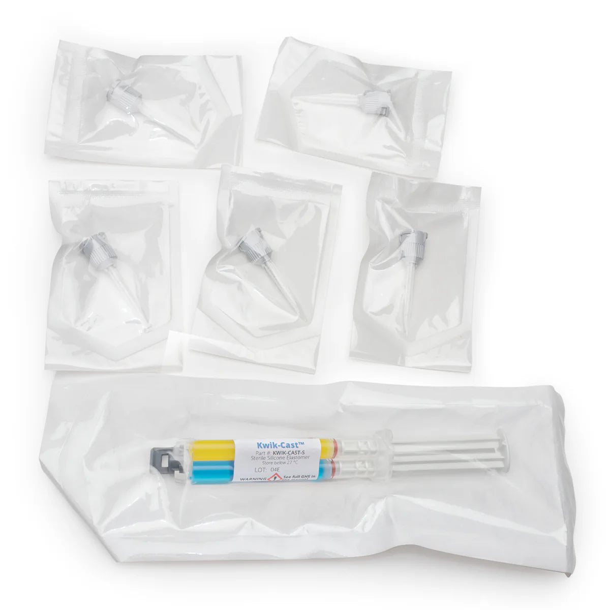
| Sterile Pkg | 1 Sterile Dual Barrel Cartridge with 5 Mixing Tips | 1 Sterile Dual Barrel Cartridge with 5 Mixing Tips |
| Best for | Chronic studies, electrode fixation | Acute recordings, nerve embedding |

Kwik-Sil™ and Kwik-Cast™ are formulated for use with live tissue, offering significantly lower toxicity than traditional RTV silicones. During curing, they release a small amount of hydrogen gas rather than acetic acid or alcohol, reducing the risk of cellular damage.
Both materials cure within minutes at room temperature without generating heat and are not inhibited by contact with biological tissue. This allows for reliable performance in applications such as electrode fixation, nerve embedding, and in vivo experimentation.
The result is a fast, reproducible silicone system that supports consistent outcomes across sensitive biological workflows.
Kwik-Sil™ is best suited for applications that require stable adhesion over time, such as chronic peripheral nerve studies and long-term electrode fixation.
Kwik-Cast™ is designed for embedding and sealing in acute recordings. It flows around tissue and electrodes to create a continuous seal that prevents fluid intrusion and supports stable signal acquisition. It can also be applied under mineral oil and allows easy electrode recovery after use.
Choose Kwik-Sil™ for adhesion and durability in longer experiments, and Kwik-Cast™ for sealing, embedding, and short-term recording setups.
Kwik-Sil™ and Kwik-Cast™ cure within minutes at room temperature, providing a short, reliable working time for precise application.
The silicone inside the cartridge cannot be sterilized by UV, gamma, H₂O₂, autoclaving, or EtO. For sterile workflows, use the pre-sterilized versions (KWIK-SIL-S™ and KWIK-CAST-S™).
The exterior of the cartridge and mixing tips can be cleaned using 70/30 alcohol or liquid sterilants such as Cidex OPA (WPI P/N 7364-4) or Rapicide OPA (WPI P/N 504611).

Kwik-Sil™ and Kwik-Cast™ come with a tip design engineered to completely mix the adhesive and minimize leakage.
NOTE: This product is non-returnable/non-refundable. For details, please refer to the WPI Terms & Conditions.
KWIK-SIL/KWIK-CAST Instruction Manual

5 Reasons KWIK-SIL & KWIK-Cast Adhesive are Ideal for Neuroscience Application
KWIK-SIL versus KWIK-Cast, What's the Difference
KWIK-SIL & KWIK-CAST: How to Install the Mixing Tip


1571


503763


VETBOND


600022

When you are looking for a low toxicity adhesive with some elasticity and good moisture resistance, choose a silicone adhesive.

Both Kwik-Sil and Kwik-Cast are silicone adhesives, but they are a little different.

The adhesive you select will depend largely on your biomedical research laboratory application. If you are building electrodes, you might use a silver-filled, conductive epoxy to connect a Ag/AgCl pellet. For instantaneous bonding when mounting a rodent brain slice, you might select an ethyl cyanoacrylate. When you are looking for a low toxicity adhesive with some elasticity and good moisture resistance, a silicone adhesive is the option of choice. When used with living tissue, an adhesive must be:

In any laboratory, having key lab supplies is almost as important as having the major equipment. Choosing a reputable supplier of these necessary supplies is as important as having quality laboratory supplies when you need them. WPI wants to be your partner in early drug discovery, and we stock a wide variety of lab supplies, many of which can ship the same business day. Having a variety of lab supplies ready to ship makes us a dependable research partner. Here are some of the popular supplies that we keep on hand to meet your needs for your upcoming experiment

If you’ve ever stepped into a research lab, you know it’s filled with specialized tools and equipment—all essential for keeping experiments running smoothly. When people ask “What are laboratory supplies” the short answer is: they’re the essentials that help scientists and researchers get their work done. That includes everything from basic items like petri dishes and pipettes to more advanced laboratory equipment like microscopes and micromanipulators.