This website uses cookies to ensure you get the best experience on our website.
Read more

Microscope Basics
April 29, 2013

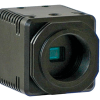
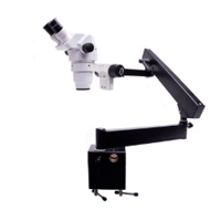
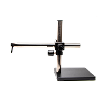
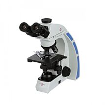
Microscopes are a standard laboratory tool, but purchasing the right microscope for a particular application can be a challenge. First, consider how you will use the instrument. Are you looking at slides, dissecting a small animal or performing a surgery? (The application dictates the necessary working distance and power of magnification.) What kind of a stand will you be using? (Boom stand, articulating arm or post stand) Will the microscope be used in a classroom setting? (A trinocular scope offers the option of including a camera.) Will you need a camera? (A camera allows you to project the microscope image on a PC or TV or to take still images.) The answers to these questions help you determine the required working distance, level of magnification, type of mounting stand and hardware required.
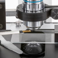
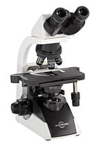
Parts of a Microscope
 Eyepiece–Two or more lenses are contained in the eyepiece, and a variety of eyepieces can be used in a microscope. Typical eyepiece magnification ranges are 10X, 15X and 20X. The eyepiece usually has a diopter adjustment for sharper focus. For information on inter-pupilary distance and adjusting the diopters, see http://micro.magnet.fsu.edu/primer/java/kohler/diopter/index.html.
Objectives–The objectives focus light from the sample. Usually three to five objectives are screwed into the nosepiece (objective turret). Objectives come in a variety of magnifications and types. For more details on the types of objectives, see "Microscope Objectives."
Stage–The stage is a platform that supports the sample. There is usually a hole in the center that allows light to pass through. The sample is positioned over the hole.
Condenser–The condenser is a lens that focuses the light from the light source onto the sample.
Light Source–While the light source could be ambient light reflected with a mirror, it is usually a halogen lamp, LED light or laser.
Focus knobs–Use the coarse and fine focus knobs to move the stage up and down to bring the sample into focus in the eyepieces.
Definitions
The following terms may be helpful when discussing microscopes:
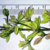
Field of View (FOV)–This is the diameter of the circle of light seen as you look through the microscope oculars. The greater the magnification, the smaller the field of view.
![]() TECH TIP: The field of view can be calculated on most compound microcopes using the field number and magnification of the objective. The FOV = field number/magnification of the objective. For example:
TECH TIP: The field of view can be calculated on most compound microcopes using the field number and magnification of the objective. The FOV = field number/magnification of the objective. For example:
18mm field number with a 10X objective --> 18/10=1.8mm=1800µm FOV.
20mm field number with a 40X objective --> 20/40=0.5mm=500µm FOV.
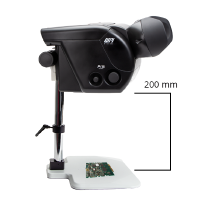
Working distance–The distance between the top of the sample and the bottom of the objective is the working distance. When dissecting a small animal, you need a greater working distance than when viewing slides.
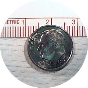
Magnification–This is the amount an image is enlarged. For example, a 10X magnification makes a sample look ten times larger. The greater the magnification, the smaller the field of view. The total magnification of a microscope equals the magnification of the eyepiece times the magnification of the objective.
Diopter–Technically, the diopter is a measure the correction value of a lens. It equals the reciprocal of the focal length in meters. If a lens can focus at 1 meter, it has a diopter of 1. If it focuses at 2 meters, it has a diopter of 1/2. Each microscope eyepiece has a diopter adjustment to allow you to make minor corrections to the image, compensating for the difference in vision between the two eyes.
Numerical aperture (N.A.)–This number is directly related to the medium of refraction (air (dry objective) = 1.00 refractive index; oil = 1.515) and the angle of refraction. An oil objective has a higher N.A. than a dry objective. The angle of refraction is determined by how close the objective is to the specimen. The higher the N.A., the greater the resolution of the objective or lens.
Resolution–The resolution describes the clarity of the view. The resolution determines how close two points can be in the field of view and still be seen as distinct.
Interpupiliary distance–This is the distance between a viewer's eyes. For binocular microscopes, you can adjust the distance between the eyepieces so that the viewer sees one circle of light instead of two when looking through both oculars.
Camera/Microscope Configuration Chart
Related Articles
-
Adjusting a Microscope
 Apr 29
 -
Choosing a Microscope Camera
 Apr 29
 -
Understanding Microscope Objectives
 Apr 29
 -
Camera Comparison Chart
 Apr 29
 -
Surgical Loupes Defining Differences
 May 01
 -
VIDEO: Digital Microforge: Setting up the Microscope and Filament
 Jan 17
 -
VIDEO: Setting up the 3D SpectaSCOPE™ - Part 1
 Feb 23
 -
How To Setup A Microscope Articulating Arm Stand
 May 27
 -
WPI's Low-Noise Amplifiers Outperform Cheap Imitations
 Apr 30
 -
How To Setup A Microscope Heavy Boom Stand
 May 27
Related Products
Recent Posts

Close